Description
By: Ming-Chon Hsiung, Wei-Hsian Yin, Fang-Chieh Lee, Wei-Hsuan Chiang
PDF: 25.4MB
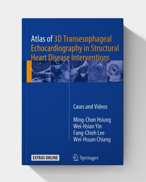
This comprehensive atlas serves as a detailed guide to the use of 3D transesophageal echocardiography (TEE) in structural heart disease interventions. The book combines high-quality images with expert insights, making it an essential resource for cardiologists, echocardiographers, and interventionalists involved in structural heart disease management.
Key Features:
- High-Quality 3D Imaging:
- Includes clear and detailed 3D TEE images that help visualize complex anatomical structures and procedural steps.
- Focuses on real-world examples to aid in the interpretation of echocardiographic findings during interventions.
- Comprehensive Coverage:
- Covers a range of structural heart disease interventions, including mitral valve repair/replacement, aortic valve procedures, and left atrial appendage occlusion.
- Offers guidance on imaging techniques, pre-procedural planning, and intra-procedural monitoring.
- Expert Insights:
- Authored by leading experts in cardiology and echocardiography, ensuring reliable and practical advice.
- Provides tips for optimizing imaging and troubleshooting common challenges during procedures.
- User-Friendly Layout:
- Organized into chapters based on different structural interventions, making it easy to navigate.
- Includes annotations and labels on images to enhance understanding.
- Practical Focus:
- Emphasizes the role of 3D TEE in improving procedural outcomes and patient safety.
- Discusses how to integrate imaging into the multidisciplinary workflow of structural heart teams.












3 reviews for Atlas of 3D Transesophageal Echocardiography in Structural Heart Disease Interventions PDF ONLY