CEDARS-SINAI • 38TH ANNUAL • 2026 EDITION
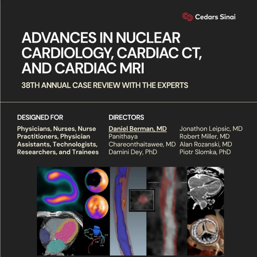
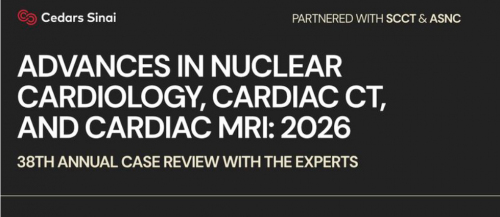
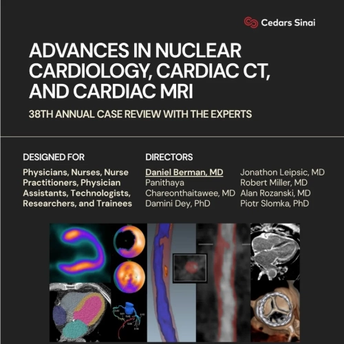
Cedars-Sinai Advances in Nuclear Cardiology, Cardiac CT and Cardiac MRI: 38th Annual Case Review with the Experts 2026
Cedars-Sinai Advances in Nuclear Cardiology, Cardiac CT and Cardiac MRI 2026 is an advanced, case-based cardiovascular imaging program focused on appropriate test selection, state-of-the-art acquisition, accurate interpretation, quantitative analysis, clinically useful reporting, and value-based application of nuclear cardiology, coronary CT angiography, PET, SPECT, and cardiac MRI.
🫀
Course Details
38th Annual Case Review with the Experts — Cedars-Sinai, Los Angeles
Original Dates: January 23–25, 2026 • Harvey Morse Conference Center, Los Angeles, CA • 4 Videos • 53 PDFs • 6.07 GB
About the Course
Cedars-Sinai Advances in Nuclear Cardiology, Cardiac CT and Cardiac MRI: 38th Annual Case Review with the Experts 2026 addresses the growing need for noninvasive cardiovascular imaging to provide measurable clinical value. Nuclear cardiology, cardiac CT, PET, SPECT, and cardiac MRI are powerful tools for diagnosis, risk assessment, and management of patients with known or suspected cardiovascular disease, but their benefit depends on selecting the right test for the right patient and performing, interpreting, reporting, and acting on each study appropriately.
The program emphasizes value-based imaging, patient safety, contemporary guidelines, quantitative imaging, multimodality decision-making, and practical case interpretation. It is designed for professionals who order, perform, interpret, or apply advanced cardiac imaging studies in routine clinical practice.
A defining feature of the course is its extensive use of case presentations and faculty discussions. The curriculum moves beyond isolated image review to show how anatomical, physiological, and quantitative findings can be integrated into clinical reports and patient-management decisions. Major 2026 themes include artificial intelligence, coronary plaque quantification, FFRct, myocardial blood-flow and flow-reserve measurement, Flurpiridaz PET, amyloid-specific tracers, photon-counting CT, structural heart planning, and advanced imaging of sarcoidosis, amyloidosis, microvascular disease, and inflammatory cardiomyopathies.
Why Choose This Cardiac Imaging Program
✓ 38th annual expert case review focused on real-world cardiovascular imaging decisions
✓ Multimodality coverage across nuclear cardiology, PET, SPECT, coronary CTA, structural CT, and cardiac MRI
✓ 4 video lectures and 53 PDF resources totaling 6.07 GB
✓ Case-based interpretation with emphasis on artifacts, pitfalls, protocol selection, and clinically meaningful reporting
✓ Advanced quantitative imaging including myocardial blood flow, flow reserve, plaque burden, calcium scoring, and disease quantification
✓ Contemporary technology including AI-assisted interpretation, FFRct, Flurpiridaz, amyloid-specific PET tracers, and photon-counting CT
✓ Patient-safety strategies to reduce unnecessary radiation and contrast exposure while maintaining high-quality studies
✓ Board-relevant knowledge review aligned with knowledge gaps commonly addressed in CBNC and CBCCT blueprint preparation
Imaging Modalities & Clinical Areas Covered
Cardiac CT: Coronary CTA • FFRct • CAC scoring • coronary plaque analysis • stents and CABG • structural heart planning • valvular disease • Watchman • TMVR • myocardial bridging • coronary anomalies • photon-counting CT
Nuclear Cardiology: PET • SPECT • MPI • Flurpiridaz • Rubidium • pharmacologic stress • stress-first protocols • amyloid imaging • PYP imaging • FDG PET • myocardial blood flow and flow reserve
Cardiac MRI: Stress MRI • perfusion • MBF-MFR • viability • amyloidosis • sarcoidosis • inflammatory cardiomyopathies • microvascular disease
Clinical Applications: Suspected and known CAD • preventive cardiology • structural heart disease • cardiac amyloidosis • cardiac sarcoidosis • ischemia • microvascular disease • valvular disease • inflammatory cardiomyopathy • PCI planning
Learning Objectives
🎯 Upon Completion You Will Be Able To
✓ Identify current evidence regarding the information provided by advanced imaging tests, appropriate test use, effects on outcomes, and costs in coronary, noncoronary, cardiac, and aortic disease.
✓ Recognize the implications of major chest-pain guidelines for the use of coronary CTA and other noninvasive testing strategies.
✓ Implement contemporary acquisition, processing, instrumentation, and software protocols across cardiac CT, PET, SPECT, and MRI.
✓ Explain how coronary flow reserve, noninvasive fractional flow reserve, plaque measurements, and other functional assessments enhance the information obtained from testing.
✓ Improve image interpretation, artifact recognition, and integration of visual and quantitative findings.
✓ Apply strategies that reduce patient radiation and contrast exposure while maintaining diagnostic study quality.
✓ Use optimal protocols for pharmacologic stress testing, including exercise-combination and pharmacologic-only approaches.
✓ Create reports that integrate test findings and provide clinically useful guidance for patient management.
✓ Apply imaging findings to management decisions using current evidence, trial data, registry findings, and guideline recommendations.
✓ Use appropriate-use criteria and evidence-based strategies to reduce unnecessary or inappropriate testing.
✓ Identify personal knowledge gaps relevant to CBNC and CBCCT board blueprints.
Complete Curriculum — Topics & Learning Themes
🧠 Section 1 — Artificial Intelligence, Quantitation & Advanced Analytics
01. AI in Cardiac CTA — Quantification of Coronary Plaque, Prediction of Risk, and Ischemia
02. Beyond Perfusion with MPI — AI Analysis of CAC, Chamber Volumes, Epicardial Fat, Body Composition and Beyond
03. Incorporating Artificial Intelligence in Interpreting Reports of SPECT MPI — Case Interpretations
04. Quantitative Plaque Analysis in Tracking Progression of Disease
05. Perfusion and Blood Flow Quantitation — Transmural and Subendocardial
🫀 Section 2 — Coronary CTA, CAD & Plaque Imaging
06. Class IA Guideline Recommendation for Chest Pain — What Evidence Brought Coronary CTA Here
07. Coronary CTA in Evaluation of Suspected CAD in Daily Practice
08. Coronary CTA in Evaluation of Patients with Known CAD — Stents and CABG
09. Coronary CTA in Asymptomatic Patients
10. Coronary CTA — Special Considerations for Women
11. Ischemia Evaluation with Coronary CTA — FFRct
12. Coronary CTA and FFRct for the Interventionalist — Use in Planning and Guiding PCI
13. Plaque Imaging in Guiding Patient Management
14. Myocardial Bridging and Coronary Anomalies on Coronary CTA — When Is Further Assessment Needed and What Should It Be?
15. Problems for Coronary CTA — Small Stents, CAC, and Breathing
16. Debate — Coronary CTA Will Replace Stress Testing
17. The Future of Cardiac CT — Photon-Counting CT and More
🧱 Section 3 — Structural Heart, Valvular Disease & Preventive CT
18. Central Role of Cardiac CT in Structural Heart Disease
19. Case Review — CTA in Valvular Disease
20. Coronary CT in Mitral Valve and Atrial Fibrillation — Watchman and TMVR
21. Coronary Artery Calcium Scanning — Central Role in Preventive Cardiology
22. Combining Anatomy and Physiology with SPECT and PET — Crucial Role of CAC Scanning in Patients
23. Evolving Trends in CAD — Shifting Risk Factors, Symptoms, and Mortality Drivers
☢️ Section 4 — PET, SPECT & Nuclear Cardiology
24. Nuclear Cardiology in 2026 and Beyond
25. PET or SPECT — Right Test for the Right Patient
26. Flurpiridaz — The Ideal PET Perfusion Tracer
27. Choosing Between Flurpiridaz and Rubidium
28. How to Start Using Flurpiridaz — Lab Preparation, Protocols, Exercise vs Pharmacologic Stress
29. Case-Based Interpretation of Flurpiridaz MPI
30. Integrating Visual and Quantitative Analyses of SPECT and PET — Improving Your Scan Interpretation with Case Illustrations
31. Stress-First SPECT Protocols — Improved Defect Detection and Stress-Only Procedures
32. Pharmacologic Stress — Combined with Exercise or Alone: What You Need to Know
📈 Section 5 — Myocardial Blood Flow, Flow Reserve & Functional Assessment
33. Clinical Value of Myocardial Blood Flow Measurement
34. How to Integrate Stress Myocardial Blood Flow and Flow Reserve in Clinical Reporting and Guiding Therapy
35. MBF-MFR with PET — How to Perform Quality Control, Interpret, and Report
36. MFR with SPECT — Ready for Prime Time?
37. MBF-MFR with Cardiac MRI — Becoming Routine in Stress MRI
38. Beyond the Images — The Essential Role of Exercise and Functional Assessment in Cardiac Testing
39. New Approaches to Functional Assessment — Coronary Flow Reserve, Noninvasive Fractional Flow Reserve, and Plaque Measurements
🧲 Section 6 — Cardiac MRI, Viability & Microvascular Disease
40. Cardiac MRI and PET in Microvascular Disease
41. Myocardial Viability — CMR and FDG PET
42. Cardiac MRI in Sarcoidosis and Other Inflammatory Cardiomyopathies
43. MRI in Cardiac Amyloidosis — Strengths and Limitations
🧬 Section 7 — Cardiac Amyloidosis
44. Amyloid Quantitation — Decreasing Subjectivity in PYP Interpretation
45. Amyloid-Specific PET Tracers
46. Tc-99m Pyrophosphate Imaging — Acquisition, Processing, Interpretation, and PET-Based Radiotracers
47. Changing the Outlook in Cardiac Amyloidosis — Current and Novel Therapies
48. The Disease, the Suspicion — Diagnostic Pathways and Treatments
🔥 Section 8 — Sarcoidosis & Inflammatory Cardiomyopathies
49. FDG PET in Sarcoidosis — Practical Considerations
50. Quantitation in Sarcoidosis Interpretation
51. Cardiac MRI in Sarcoidosis and Other Inflammatory Cardiomyopathies
🎓 Section 9 — Expert Case Review, Reporting & Value-Based Imaging
52. My Best Teaching Cases
53. Putting It All Together — Value-Based Use of Cardiac Imaging
54. Game-Show Jeopardy — Cardiac CT, Nuclear, and CMR
55. Creating Optimal Reports That Integrate Test Findings and Guide Patient Management
56. Using Criteria and Evidence-Based Strategies to Reduce Inappropriate Testing
57. Board Blueprint Review — Identifying Knowledge Gaps for CBNC and CBCCT Examinations
Curriculum note: The sections above preserve the supplied session topics and also incorporate key learning themes from the stated objectives. The course uses cases and expert discussion to connect image acquisition and interpretation with reporting, appropriate test selection, patient safety, costs, clinical outcomes, and patient-management decisions.
Who Should Take This Course
Perfect for: cardiologists and radiologists who order, perform, interpret, report, or apply advanced cardiovascular imaging studies.
✓ Cardiologists ✓ Cardiac imaging specialists ✓ Cardiac radiologists ✓ General radiologists
✓ Nuclear medicine physicians ✓ Nuclear cardiologists ✓ Interventional cardiologists ✓ Preventive cardiologists
✓ Cardiology and radiology fellows ✓ Physicians preparing for CBNC or CBCCT knowledge review
✓ Nurse practitioners ✓ Physician assistants ✓ Radiologic and nuclear medicine technologists
✓ Imaging researchers, technicians, and professionals supporting cardiac CT, PET, SPECT, or MRI laboratories
Course Format & Original Event Information
📥 4 Videos + 53 PDFs
The educational package contains four video files and 53 PDF resources, with a total size of approximately 6.07 GB.
📅 January 23–25, 2026
Originally scheduled from Friday, January 23, 2026 at 7:30 AM through Sunday, January 25, 2026 at 2:55 PM Pacific Time.
📍 Cedars-Sinai, Los Angeles
The original live activity was held at the Cedars-Sinai Harvey Morse Conference Center in Los Angeles, California.
⚠️ Educational Content
This listing describes digital educational materials. Conference attendance, certification, examination registration, and CME credit are not stated as included.



Reviews
There are no reviews yet.